Photo Gallery
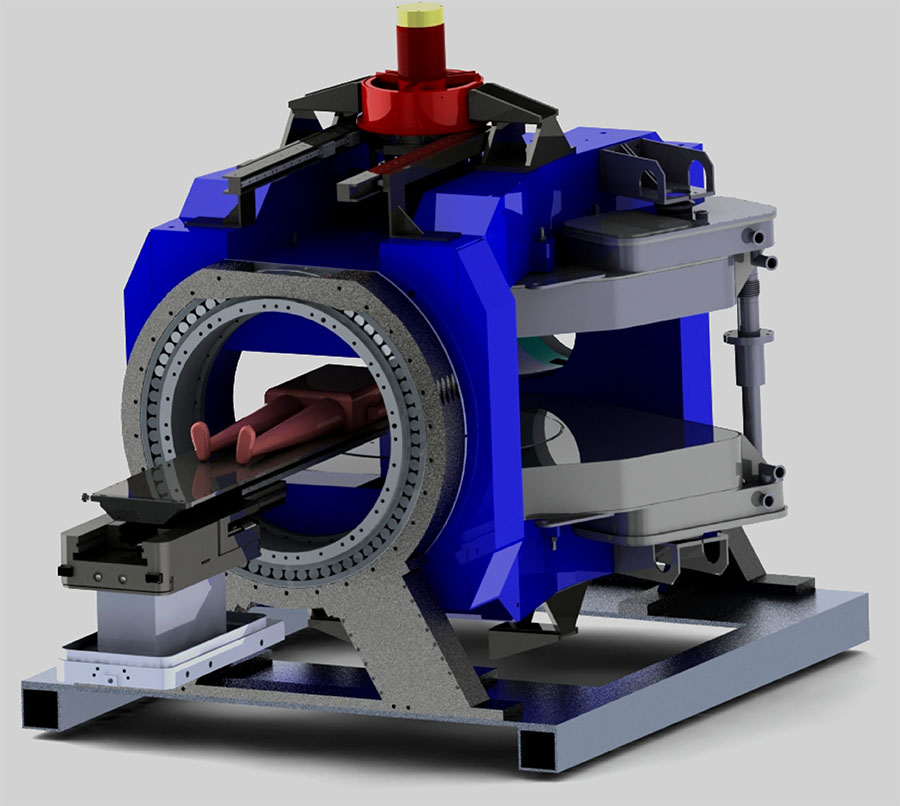
Clinical Whole Body Linac-MR v.2
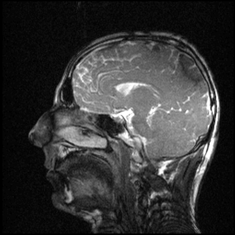
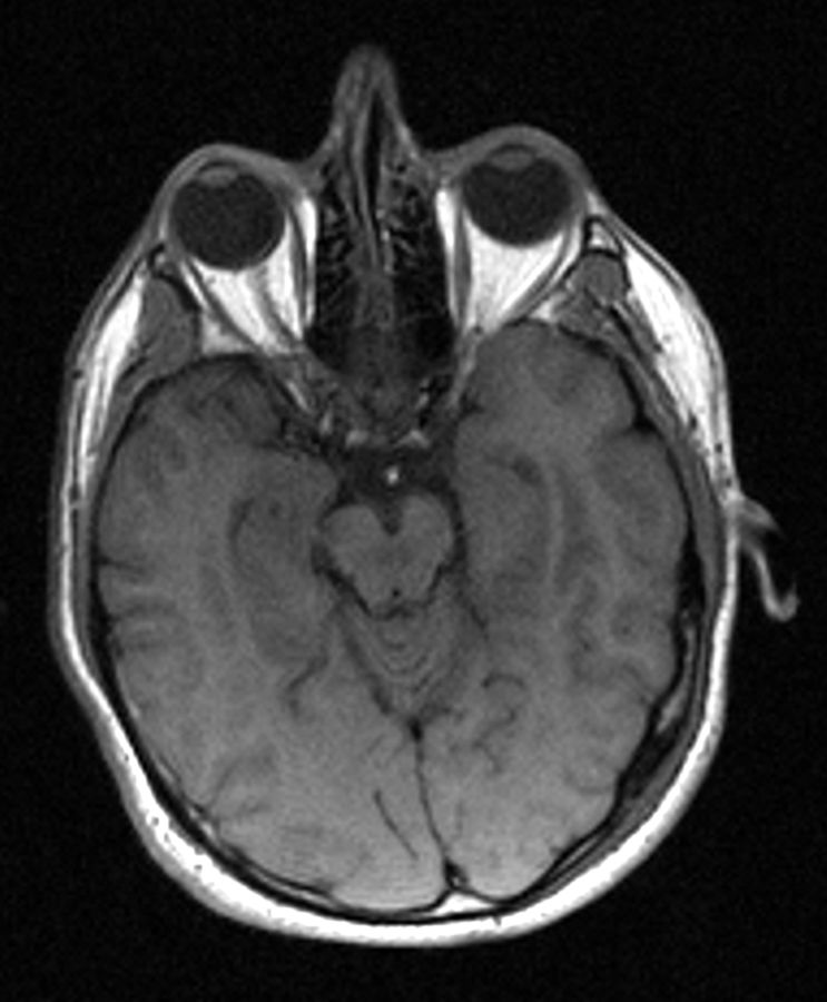
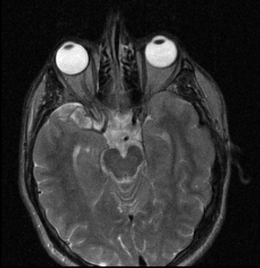
First Linac-MR images of a Human Subject
The Whole Body Linac-MR v.1 imaged it's first human subject in July 2014 producing these sample images.
Clinical Whole Body Linac-MR v.1 installed in 2013
6 MV Linac; 0.5 T MR; 60 cm bore
The Whole Body Linac-MR v.1 is installed into an existing clinical vault (depth: 19.4 ft., width: 19.8 ft., height: 12 ft.) without removing and rebuilding the walls or ceiling. The MR magnet features a high temperature superconducting magnet that does not require liquid helium or a helium exhaust vent.
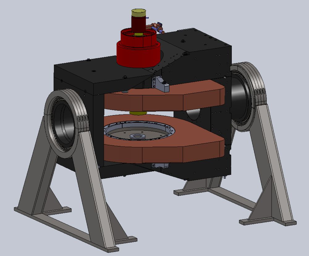
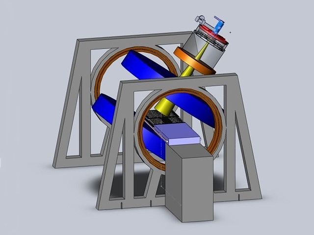
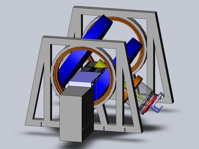
Models of Whole Body Linac-MR v.1 Design
Transverse (Perpendicular) Configuration
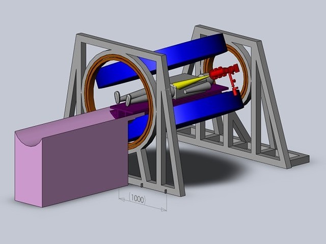
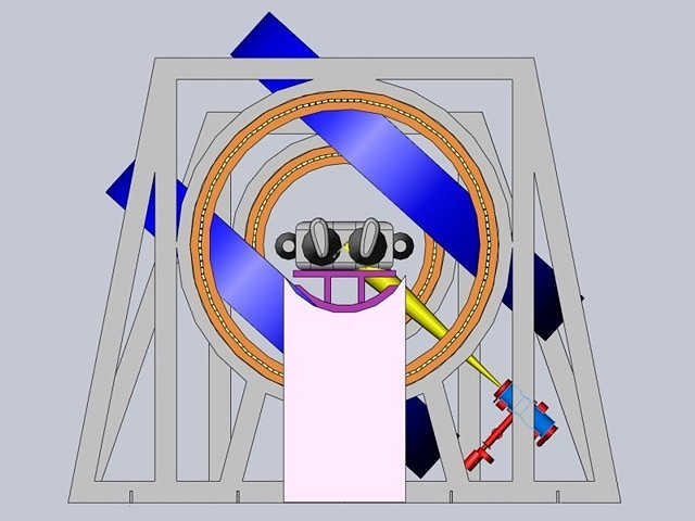
Longitudinal (Parallel) Configuration
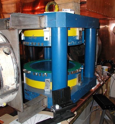
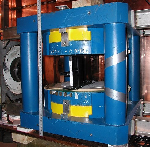
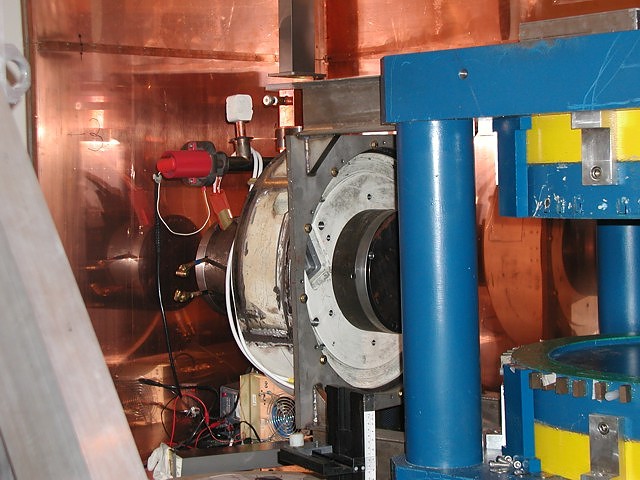
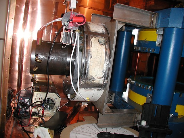
First Images Obtained in 2008 from the Head Prototype
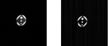
Axial slices were obtained with a raw gradient echo sequence, flip angle =90o, TE=14.0 ms, TR=300 ms, Bandwidth = 10 kHz, matrix size=128x128, No-Averages=1, slice thickness=7 mm, FOV=100 mm x 100 mm. The phantom consists of an acrylic rectangular cube 15.95 x 15.95 x 25.4 mm (0.628 x 0.628 x 1.0 inch) with holes of diameter 2.52 mm, 3.45 mm and 4.78 mm (0.099 inch, 0.136 inch, 0.188 inch) drilled parallel to the length of the rectangular cube. The cube is immersed in a 10 mM solution of CuSO4 within a plastic container 22.5 mm inner diameter.
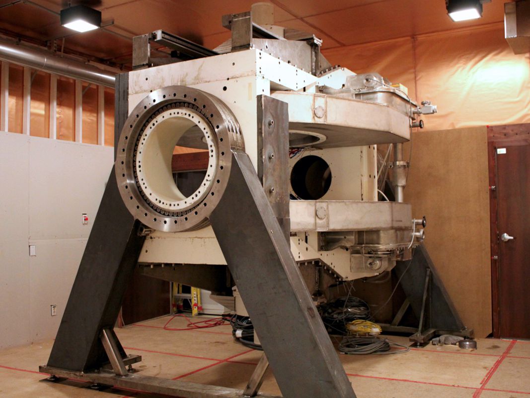
Prototype Head Linac-MR (during Construction)