Real-time MR-Guided Radiotherapy
Imagine trying to aim at a moving target, knowing that if you hit outside the bull’s eye, the consequences could be serious. In this case, the bull’s eye is a tumor and missing the mark could mean serious side effects for patients receiving radiation therapy.
Radiation treatment is a proven cancer treatment. Yet, targeting the radiation beam on organs that move—the lungs, liver, stomach and pancreas, for instance—can be difficult. But Cancer Care Alberta - Alberta Health Services (ACC-AHS) researcher Dr. Gino Fallone is hoping it won’t be long before the mark becomes easier to hit.
Dr. Fallone is leading a team to develop a radiation therapy MRI system that will use a process called Advanced Real-Time Adaptive Radiotherapy (ART)2. For years, people have been trying to pair an MRI machine with a linear accelerator to improve the accuracy of radiation beams for solid tumors. But Dr. Fallone’s team will be the first to get it done.
“These two machines are allergic to each other,” says Dr. Fallone. “The radio frequency and magnetic fields from each are not compatible with the other. We have completely redesigned the system from “the bottom-up” and have resolved these issues that have troubled people for years.”
The hybrid system, developed by Dr. Fallone and Brad Murray, also an ACC-AHS researcher, will allow clinicians to administer radiation at the same time “exquisite imaging” is taking place.
Currently, when a patient is treated with radiation therapy, a medical team studies the patient’s tumor through a set of images—x-rays, ultrasounds, MRI, or CTs. They must then use a complex calculation process to map where radiation should be aimed. Often days pass between the first set of images and the actual administration of radiation, which means the tumor’s shape and location may have changed. Since it is difficult to know exactly where the tumor is, doctors will treat a small area of potentially healthy tissue around the cancer to ensure they are reaching the whole tumor. Damaging this healthy tissue is the cost of not knowing the exact location of the tumor during radiation treatments.
There are so called “work around” techniques used to improve accuracy, but they can be invasive and inexact, says Dr. Fallone. With the hybrid system, high-quality real-time 3 D images are available at the time of treatment irradiation. In other words, the medical team can capture a precise picture of the tumor and treat it immediately.
“Without this type of technology, you have to decrease the dose of the radiation because some radiation will hit healthy tissue,” says Dr. Fallone. “Because this new system will allow us to precisely place the radiation beam on the patient’s tumor, the side effects will be reduced and we don’t have to worry about decreasing the dose. In other words, we can cure the cancer much better and without side effects.”
Aside from improving the accuracy of radiation treatments for solid tumors, it will mean certain types of tumors that are not normally treated by radiation now—liver, stomach and pancreatic cancers—can include radiation as treatment option. As well, outcomes for lung and prostate cancers should improve since it is currently difficult to administer enough radiation to properly treat the tumor.
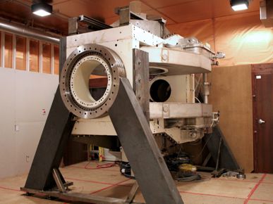
The whole body prototype began installation in 2013. The first Linac-MR images on a human volunteer were obtained in July 2014. Testing of the radiation treatment component is progressing. See other pictures in the gallery.